Founded in 1846 in Jena, ZEISS is headquartered in Oberkochen, Germany, and continues to be a leader in innovation and precision technologies.
ZEISS is a globally renowned technology enterprise specializing in optics and optoelectronics. The company provides advanced microscopy solutions for life sciences and materials research, serving a broad spectrum of scientific and industrial customers.
With a workforce of over 35,000 employees, ZEISS operates in nearly 50 countries, encompassing approximately 30 production facilities, 60 sales and service locations, and 27 research and development centers.
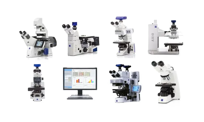
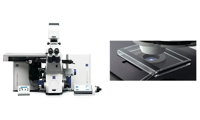
Upright and Inverted Widefield Microscopes
Widefield microscopes are offered in various optical detection configurations. An upright microscope is ideal for analyzing zebrafish embryos, stained tissue sections, or brain slices. For tissue culture applications and rapid evaluations in routine research, an inverted microscope may be the more suitable option.
Key Features
- Axio – Upright and inverted models for life science research and materials microscopy
- Axioscope – Smart microscope designed for biomedical routine tasks, research, and materials analysis
- Axiolab – Optimized for daily laboratory operations
- Axio Imager Vario – Upright microscope built for examining large samples
- Axio Imager – Versatile imaging microscope for detailed analysis
- Primo Series – Upright and inverted microscopes for digital teaching and routine lab work
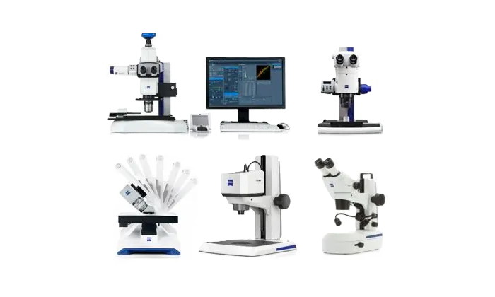
Stereo and Zoom Microscopes
Widefield microscopes are offered in various optical detection configurations. An upright microscope is ideal for analyzing zebrafish embryos, stained tissue sections, or brain slices. For tissue culture applications and rapid evaluations in routine research, an inverted microscope may be the more suitable option.
Key Features
- Axio – Upright and inverted models for life science research and materials microscopy
- Axioscope – Smart microscope designed for biomedical routine tasks, research, and materials analysis
- Axiolab – Optimized for daily laboratory operations
- Axio Imager Vario – Upright microscope built for examining large samples
- Axio Imager – Versatile imaging microscope for detailed analysis
- Primo Series – Upright and inverted microscopes for digital teaching and routine lab work
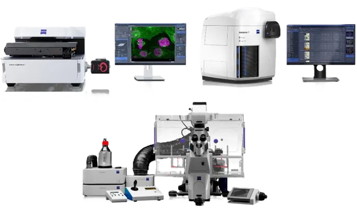
Imaging Systems
Imaging systems integrate traditional wide-field microscopy with advanced computing and imaging technologies. These solutions are ideal for highly automated workflows and in-depth subcellular research.
Key Features
- Lattice Lightsheet – Enables long-term, high-resolution volume imaging of live cells
- Axioscan – Designed for high-throughput slide scanning in biology and geosciences
- Cell Observer – Allows real-time visualization of live cells and organisms
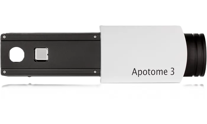
Apotome 3
Create precise optical sections of your fluorescent samples with structured illumination. ZEISS Apotome 3 simplifies the removal of out-of-focus light, allowing you to concentrate fully on your research. The system automatically recognizes magnification levels and positions the correct grid into the beampath. Optical sections are then generated from multiple images using different grid phases, delivering consistently reliable results—even in thick specimens. Despite its advanced capabilities, the system remains user-friendly and intuitive. Expect high-contrast images with exceptional resolution—brilliant optical sections made simple.
Key Features
- Brilliant Optical Sections: Equipped with three grids of varying geometries, Apotome 3 ensures optimal resolution at any magnification.
- Flexible Light Source and Dye Compatibility: Compatible with a wide range of fluorophores and light sources, allowing flexibility as experimental complexity increases.
- Enhanced Structural Detail: Advanced deconvolution using a patented structured illumination algorithm provides improved visualization of fine structural details.
Laser Microdissection and Optical Tweezers
Laser Microdissection offers a contamination-free solution for isolating specific cells from tissue samples or cell cultures. This precise separation method provides pure cell populations, ideal for producing accurate and reliable molecular analysis results.
Key Features
- Fully integrated system combining laser microdissection with imaging workstation capabilities
- Compatible with standard glass slides, including archived materials, using ZEISS-patented sample capture technology
- Expandable platform with AxioCam digital camera options for fluorescence, brightfield, multi-channel fluorescence, and extended focus imaging
- Flexible integration of additional components and customizable workflows, suitable for both individual and automated applications
- Calibration routines allow easy adjustment of laser parameters based on specimen characteristics
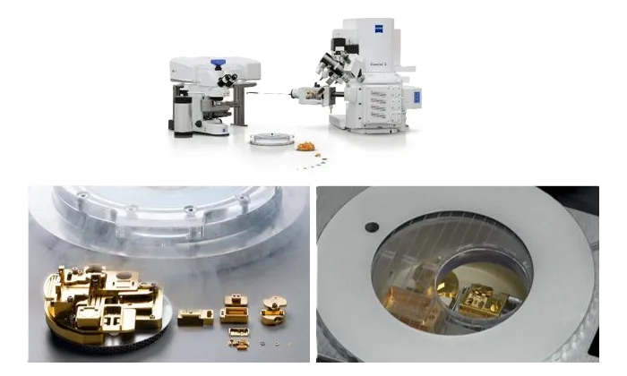
ZEISS Correlative Cryo Workflow
Cryogenic microscopy enables the observation of cellular structures in a near-native state by instantly halting biological processes and preserving ultrastructure without artifacts.
The ZEISS Correlative Cryo Workflow integrates widefield, laser scanning, and focused ion beam scanning electron microscopy into a streamlined, user-friendly process. This solution includes both hardware and software tailored for correlative cryogenic workflows—supporting everything from fluorescent macromolecule localization to high-contrast 3D imaging and precise on-grid lamella thinning for cryo-electron tomography.
Key Features
- Seamless cryogenic workflow across widefield, confocal, and FIB-SEM modalities
- Effective protection against devitrification and ice contamination
- High-resolution cryo-fluorescence imaging
- High-contrast volume imaging and 3D reconstruction
- Targeted on-grid lamella thinning for cryo-TEM applications
- Versatile use for both cryogenic and ambient conditions
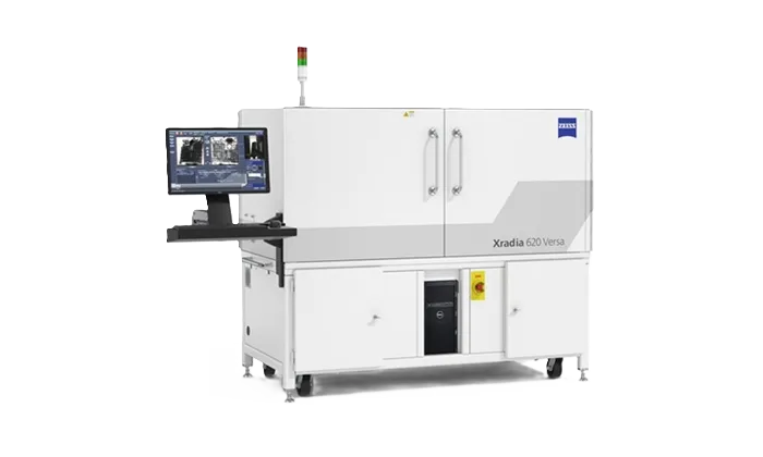
3D X-Ray
Non-Destructive 3D X-Ray Microscopy (XRM) for University, R&D Center, Semiconductor Failure Analysis, Advanced Packaging, Materials Science, Life Sciences Applications.