


 繁體中文
繁體中文 English
ZEISS


ZEISS is an internationally leading technology enterprise operating in the fields of optics and optoelectronics. For its customers, ZEISS develops microscopy solutions for the life sciences and materials research. With over 35,000 employees, ZEISS is active globally in almost 50 countries with around 30 production sites, 60 sales and service companies and 27 research and development facilities. Founded in 1846 in Jena, the company is headquartered in Oberkochen, Germany.
Upright and Inverted Widefield Microscopes
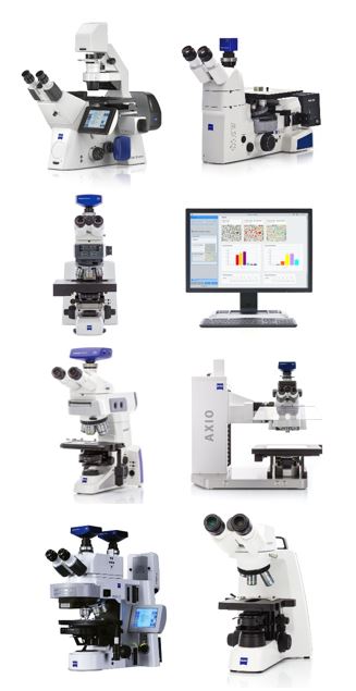
Widefield microscopes are available in different optical detection versions. Choose an upright microscope if your task is to analyze zebrafish embryos, stained tissue selections or brain slices. For tissue culture and quick assessments in routine research, the inverted microscope might be the better choice.
Main features
Axio - Upright/Inverted Life Science Research, Materials Microscopy
Axioscope - Smart microscope for biomedical routine, research, materials
Axiolab - designed for the day-to-day work performed in the laboratory
Axio Imager Vario - Upright microscope for large samples
Axio Imager - Imager
Primo-Series Upright/Inverted Microscopes for Digital Teaching and Routine Laboratory Work
Application
Applications in the fields of biology, materials, research, etc., are tailored to your optical detection application and can be flexibly expanded as needed.
Stereo and Zoom Microscopes
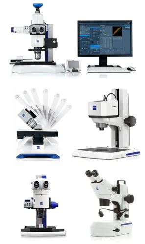
Generate three-dimensional and laterally precise images. With a Stereo Microscope, you observe large samples such as leaves and tissues or inspect rough material surfaces. Upgrade your microscope flexibly with different optical detection applications in the fields of biology, materials, research, etc., which are tailored to your optical detection application and can be flexibly expanded as needed. digital cameras and benefit from various types of illumination techniques.
Main features
Axio Zoom.V16 - 2013 Red Dot Design Award Winner, Large Field of View (Fluorescence) Stereo Zoom Microscope
Smartzoom - 2015 Red Dot Design Award Winner for Automated Digital Zoom Microscopes for Routine and Failure Analysis
Visioner Series-Full Focus Optical Inspection Microscope
SteREO Series - Zoom Microscope
Stemi Series - Stereo Microscope
Application
Large field of view, extended working distance, and flexible expansion for Living Cells, Organisms, Uniquely designed for petrographic optical detection analysis applications in biology, materials, research, and other fields.
Imaging Systems
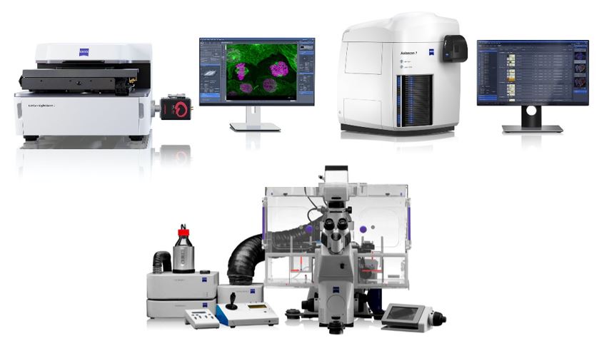
Imaging Systems mostly combine traditional wide-field microscopy components with powerful computer hardware and sophisticated imaging technologies. These products are the right solutions for your highly automated tasks and subcellular investigations.
Main features
Lattice Lightsheet - Long-Term Volume Imaging of Live Cells
Axioscan - Biology, Geosciences
Cell Observer - Visualize living organisms and cells
Application
Living Cells, Organisms, Uniquely designed for petrographic analysis
ZEISS Apotome 3
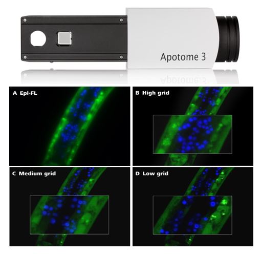
Create optical sections of your fluorescent samples - with structured Illumination, removal of out-of-focus light becomes simple and efficient, allowing you to fully focus on your research. ZEISS Apotome 3 recognizes the magnification and moves the appropriate grid into the beampath. The system then calculates your optical section from a number of images with different grid positions. It’s a totally reliable way to remove out-of-focus light, even in thicker specimens. Yet your system remains just as easy to operate as always. You get images with high contrast in the best possible resolution – simply brilliant optical sections.
Main features
Brilliant Optical Sections: Apotome 3 comes with three grids of different geometries, giving you the best resolution no matter which magnification you choose.
Free Choice of Light Source and Dyes: Apotome 3 adapts to your fluorophores and lights source. So you stay flexible when your experiments evolve in complexity and requirements.
More Structural Information: Your created images are improved even more by deconvolution, using a patented algorithm for structured illumination. Better recognize important structures of your examined objects.
Application
Living Cells, Organisms, Uniquely designed for petrographic optical detection analysis.
Laser Microdissection and Optical Tweezers
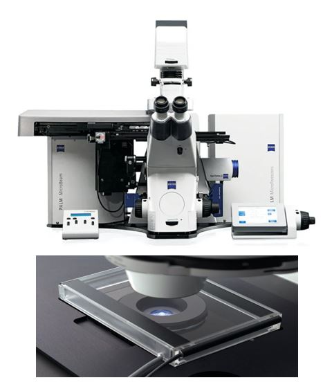
Laser Microdissection is the contamination-free method for separating and collecting cells of interest out of tissue samples or cell cultures. The isolated pure cell populations are the ideal starting point for specific and meaningful results in your molecular analysis.
Main features
A system that completes the step from laser-microdissection to integrated imaging workstation.
Performing LCM even from standard glass slides, e.g. archived material, with ZEISS-patented sample capture technology.
Extendibility – add digital cameras from the AxioCam camera range for fluorescence, bright field, multi-channel fluorescence and extended focus.
Ability to integrate additional components into MicroBeam and create simple workflows, whether for individual experiments or automation with flexible collectors.
Calibration routines that allow you to easily adjust MicroBeam for laser parameters appropriate to your chosen specimen.
Application
Suitable for laser microdissection and analysis for DNA, RNA and protein isolation — whether from archive material or live cells.
Applicable to cryosections, FFPE (formalin fixed and paraffin embedded) tissue.
ZEISS Correlative Cryo Workflow
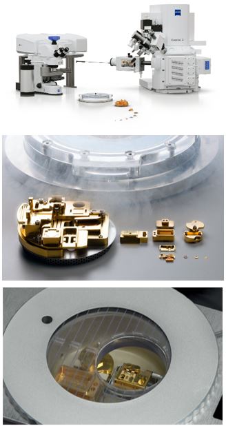
Cryogenic microscopy allows the examination of cellular structures in their near-to-native state, as the ultrastructure of cells and tissues can be preserved free of artifacts and cellular processes are stopped instantaneously.
ZEISS Correlative Cryo Workflow connects widefield, laser scanning, and focused ion beam scanning electron microscopy in a seamless and easy-to-use procedure. The solution provides hardware and software optimized for the needs of correlative cryogenic workflows, from localization of fluorescent macromolecules to high-contrast volume imaging and on-grid lamella thinning for cryo electron tomography.
Main features
Seamless cryogenic workflow across multiple modalities
Sample protection against devitrification and ice contamination
High-resolution fluorescence imaging
High-contrast volume imaging and 3D reconstruction
Targeted on-grid lamella thinning for cryo TEM applications
Multipurpose use for cryogenic and room temperature applications
Application
Cell Biology
Cancer research
Plant science
Developmental Biology